
Page 395 - Atlas of Small Animal CT and MRI
P. 395
Brachial and Lumbosacral Plexus 385
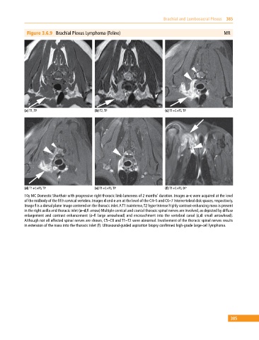
Figure 3.6.9 Brachial Plexus Lymphoma (Feline) MR
(a) T1, TP (b) T2, TP (c) T1+C+FS, TP
(d) T1+C+FS, TP (e) T1+C+FS, TP (f) T1+C+FS, DP
10y MC Domestic Shorthair with progressive right thoracic limb lameness of 2 months’ duration. Images a–c were acquired at the level
of the midbody of the fifth cervical vertebra. Images d and e are at the level of the C4–5 and C6–7 intervertebral disk spaces, respectively.
Image f is a dorsal plane image centered on the thoracic inlet. A T1 isointense, T2 hyperintense highly contrast‐enhancing mass is present
in the right axilla and thoracic inlet (a–d,f: arrow) Multiple cervical and cranial thoracic spinal nerves are involved, as depicted by diffuse
enlargement and contrast enhancement (c–f: large arrowhead) and encroachment into the vertebral canal (c,d: small arrowhead).
Although not all affected spinal nerves are shown, C5–C8 and T1–T2 were abnormal. Involvement of the thoracic spinal nerves results
in extension of the mass into the thoracic inlet (f). Ultrasound‐guided aspiration biopsy confirmed high‐grade large‐cell lymphoma.
385