
Page 394 - Atlas of Small Animal CT and MRI
P. 394
384 Atlas of Small Animal CT and MRI
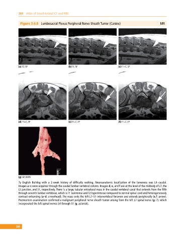
Figure 3.6.8 Lumbosacral Plexus Peripheral Nerve Sheath Tumor (Canine) MR
(a) T2, SP (b) T1, SP (c) T1+C, SP
(d) T1+C, TP (e) T1+C, TP (f) T1+C, TP
(g) GP, DORS
7y English Bulldog with a 2‐week history of difficulty walking. Neuroanatomic localization of the lameness was L4–caudal.
Images a–c were acquired through the caudal lumbar vertebral column. Images d, e, and f are at the level of the midbody of L7, the
LS junction, and S1, respectively. There is a large, tubular extradural mass in the caudal vertebral canal that extends from the fifth
through seventh lumbar vertebrae, which is T1 isointense and T2 hyperintense compared to normal spinal cord and heterogeneously
contrast enhancing (a–d: arrowhead). The mass exits the left L7–S1 intervertebral foramen and extends peripherally (e,f: arrow).
Postmortem examination confirmed a malignant peripheral nerve sheath tumor arising from the left L7 spinal nerve (g: 7), which
incorporated the left spinal nerves L4 through S1 (g: asterisk).
384