
Page 401 - Atlas of Small Animal CT and MRI
P. 401
Thoracic wall and Diaphragm 391
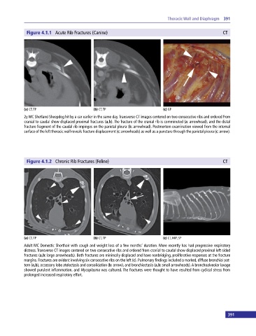
Figure 4.1.1 Acute Rib Fractures (Canine) CT
(a) CT, TP (b) CT, TP (c) GP
2y MC Shetland Sheepdog hit by a car earlier in the same day. Transverse CT images centered on two consecutive ribs and ordered from
cranial to caudal show displaced proximal fractures (a,b). The fracture of the cranial rib is comminuted (a: arrowhead), and the distal
fracture fragment of the caudal rib impinges on the parietal pleura (b: arrowhead). Postmortem examination viewed from the internal
surface of the left thoracic wall reveals fracture displacement (c: arrowheads) as well as a puncture through the parietal pleura (c: arrow).
Figure 4.1.2 Chronic Rib Fractures (Feline) CT
(a) CT, TP (b) CT, TP (c) CT, MIP, SP
Adult MC Domestic Shorthair with cough and weight loss of a few months’ duration. More recently has had progressive respiratory
distress. Transverse CT images centered on two consecutive ribs and ordered from cranial to caudal show displaced proximal left‐sided
fractures (a,b: large arrowheads). Both fractures are minimally displaced and have nonbridging, proliferative responses at the fracture
margins. Fractures are evident involving six consecutive ribs on the left (c). Pulmonary findings included a marked, diffuse bronchial pat-
tern (a,b), accessory lobe atelectasis and consolidation (b: arrow), and bronchiectasis (a,b: small arrowheads). A bronchoalveolar lavage
showed purulent inflammation, and Mycoplasma was cultured. The fractures were thought to have resulted from cyclical stress from
prolonged increased respiratory effort.
390 391