Page 405 - Atlas of Small Animal CT and MRI
P. 405

Thoracic wall and Diaphragm  395


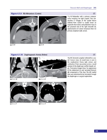
              Figure 4.1.9  Rib Metastasis (Canine)                                                       CT
                                                                             11y M Rottweiler with a primary osteosar-
                                                                             coma involving the right scapula. Two con-
                                                                             secutive CT images of the caudal thorax
                                                                             ordered from cranial to caudal reveal an
                                                                             expansile destructive and productive mass in
                                                                             the proximal end of the right eleventh rib,
                                                                             representative of a bone metastasis from the
                                                                             primary neoplasm (a,b: arrow).


            (a) CT, TP                       (b) CT, TP


              Figure 4.1.10  Diaphragmatic Hernia (Feline)                                                CT
                                                                             10y MC Domestic Longhair referred for a cau-
                                                                             dal thoracic mass. An ovoid mass is seen in
                                                                             the  caudoventral  thorax (a,b: arrow), and
                                                                             continuity of liver vasculature across a small
                                                                             window in the diaphragm verifies hepatic ori-
                                                                             gin. Transverse images show the appearance
                                                                             of the abdominal (c) and herniated thoracic
                                                                             (d) components of the liver. The left medial
                                                                             lobe was determined to be herniated through
                                                                             the diaphragm on surgical exploration.
            (a) CT+C, DP                     (b) CT+C, SP


            (c) CT+C, TP                     (d) CT+C, TP


 394                                                                                                         395
   400   401   402   403   404   405   406   407   408   409   410