
Page 410 - Atlas of Small Animal CT and MRI
P. 410
400 Atlas of Small Animal CT and MRI
or encroach on the pleura. Mesotheliomas may appear Pleural thickening/fibrosis
as a discrete mass on CT images but can go undetected
in patients in which the tumor invades the pleura dif- Pleural thickening is sometimes evident without other
fusely (Figure 4.2.10). CT imaging features of other associated radiographic abnormalities or clinical signs
8,9
masses depend on anatomic location and tumor type and is thought to be due to previous pleural inflamma-
(Figure 4.2.11). 9 tory disease with resulting pleural fibrosis (Figure 4.2.12). 9
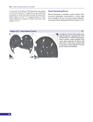
Figure 4.2.1 Pneumothorax (Canine) CT
12y Doberman Pinscher with an axillary mass.
Image b represents a magnified view of the
left dorsal thorax. A mild left‐sided pneumo-
thorax is present, causing retraction of the
visceral pleura away from the thoracic wall
(a,b: arrows). A small volume of pleural fluid
is also present, causing separation of visceral
pleural surfaces (a: arrowheads).
(a) CT, TP (b) CT, TP
400 401