
Page 414 - Atlas of Small Animal CT and MRI
P. 414
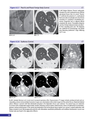
Figure 4.2.7  Pleuritis and Pleural Foreign Body (Canine)                                    CT

                                                                              2y MC Belgian Malinois. Thoracic radiographs
                                                                              revealed a focal left caudal pulmonary  infiltrate
                                                                              and  pleural  fissure lines  (a:  arrow). Thoracic
                                                                              CT confirmed the presence of a small pleural
                                                                              fluid volume (b: arrowheads) and a focal lesion
                                                                                consisting of a peripheral consolidating pul-
                                                                              monary component and an adjacent pleural
                                                                              component (b: arrow). Presumptive diagnosis
                                                                              was focal foreign‐body pneumonia and pleuri-
                                                                              tis from migrating plant awn. The diagnosis
                                                                              was  confirmed  by  bronchoscopy  and  partial
                                                                              lung lobectomy performed 3 days   following
                                                                              the CT scan.

             (a) DX, LAT                      (b) CT, TP


              Figure 4.2.8  Pyothorax (Canine)                                                             CT


             (a) CT, TP                       (b) CT, TP                       (c) CT+C, DP


             (d) CT+C, TP                     (e) CT+C, TP
             8y MC Labrador Retriever with recent‐onset increased respiratory effort. Representative CT images include unenhanced (a,b) and cor-
             responding contrast‐enhanced (d,e) transverse images and a dorsal plane reformatted image (c) of the ventral thorax. Moderate bilateral
             pleural effusion is present with fluid distributed primarily in the dependent thorax (a,b: asterisks). The ventral lungs are atelectatic
             (a: arrows) with nondependent regions better aerated. Following contrast medium administration, there is ill‐defined but marked hetero-
             geneous contrast enhancement in the ventral paramediastinal and ventral pleural space regions (c,e: arrows). Surgical exploration with
             biopsy revealed chronic fibrinosuppurative pleuritis with villonodular mesothelial proliferation and multifocal abscessation. A plant awn
             foreign body was found in the suppurative effusion.


           404                                                                                                                                                                                                                       405