
Page 415 - Atlas of Small Animal CT and MRI
P. 415
Pleural Space 405
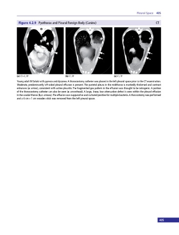
Figure 4.2.9 Pyothorax and Pleural Foreign Body (Canine) CT
(a) CT+C, TP (b) CT, TP (c) CT, TP
Young adult M Saluki with pyrexia and dyspnea. A thoracostomy catheter was placed in the left pleural space prior to the CT examination.
Moderate, predominantly left‐sided pleural effusion is present. The parietal pleura in the midthorax is markedly thickened and contrast
enhances (a: arrow), consistent with active pleuritis. The fragmented gas pattern in the effusion was thought to be iatrogenic. A portion
of the thoracostomy catheter can also be seen (a: arrowhead). A large, linear, low‐attenuation defect is seen within the pleural effusion
in the caudal thorax (b,c: arrows). The effusion was suppurative and cultured positive for multiple bacteria. A thoracotomy was performed
and a 6 cm × 1 cm wooden stick was removed from the left pleural space.
404 405