
Page 420 - Atlas of Small Animal CT and MRI
P. 420
410 Atlas of Small Animal CT and MRI
arising in the mediastinum. The esophagus appears as Esophageal neoplasia
a thin soft‐tissue attenuating crescent associated with Neoplasia of the esophagus is rare and includes carci-
part of the abscess margin on transverse CT images noma, sarcoma (associated with Spirocerca lupi infec-
(Figure 4.3.16). Abscesses are fluid attenuating on unen- tion), leiomyoma, leiomyosarcoma, and lymphoma.
hanced CT images and peripherally contrast enhance. Imaging features depend on the size and location of
The flattened esophageal mucosa has a characteristic the mass. Obstruction may be a sequela with resulting
curvilinear pattern of contrast enhancement conform- esophageal dilation cranial to site of the neoplasm.
ing to the curvature of the abscess. Additional imaging Neoplasms may be solid or heterogeneous on CT and
features associated with mediastinitis may also be pre- MR images and typically appear as an eccentric or cir-
sent. The adjacent lung lobes can sometimes be atelec- cumferential mass. The intensity and pattern of contrast
tatic as a result of encroachment by the mass. 8 enhancement are variable (Figure 4.3.17).
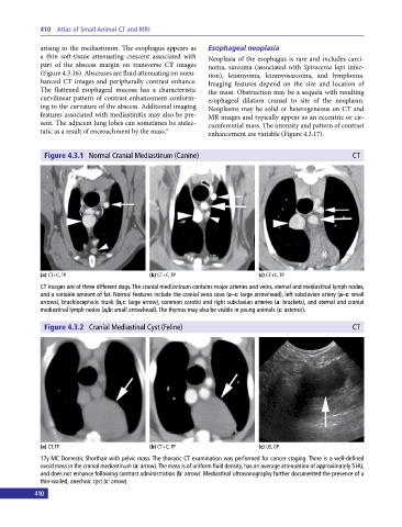
Figure 4.3.1 Normal Cranial Mediastinum (Canine) CT
(a) CT+C, TP (b) CT+C, TP (c) CT+C, TP
CT images are of three different dogs. The cranial mediastinum contains major arteries and veins, sternal and mediastinal lymph nodes,
and a variable amount of fat. Normal features include the cranial vena cava (a–c: large arrowhead), left subclavian artery (a–c: small
arrows), brachiocephalic trunk (b,c: large arrow), common carotid and right subclavian arteries (a: brackets), and sternal and cranial
mediastinal lymph nodes (a,b: small arrowhead). The thymus may also be visible in young animals (c: asterisk).
Figure 4.3.2 Cranial Mediastinal Cyst (Feline) CT
(a) CT, TP (b) CT+C, TP (c) US, OP
17y MC Domestic Shorthair with pelvic mass. The thoracic CT examination was performed for cancer staging. There is a well‐defined
ovoid mass in the cranial mediastinum (a: arrow). The mass is of uniform fluid density, has an average attenuation of approximately 5 HU,
and does not enhance following contrast administration (b: arrow). Mediastinal ultrasonography further documented the presence of a
thin‐walled, anechoic cyst (c: arrow).
410