Page 422 - Atlas of Small Animal CT and MRI
P. 422

412  Atlas of Small Animal CT and MRI


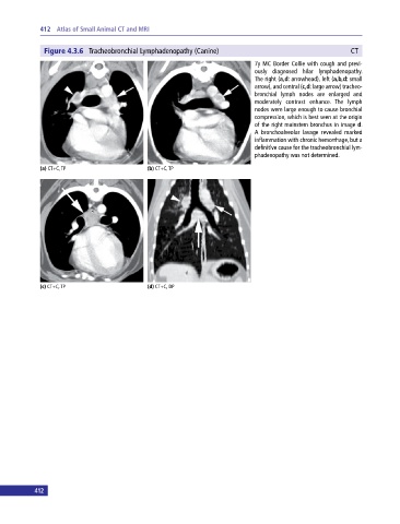
              Figure 4.3.6  Tracheobronchial Lymphadenopathy (Canine)                                      CT
                                                                              7y MC Border Collie with cough and previ­
                                                                              ously diagnosed hilar lymphadenopathy.
                                                                              The right (a,d: arrowhead), left (a,b,d: small
                                                                              arrow), and central (c,d: large arrow) tracheo­
                                                                              bronchial lymph nodes are enlarged and
                                                                              moderately contrast enhance.  The lymph
                                                                              nodes were large enough to cause bronchial
                                                                              compression, which is best seen at the origin
                                                                              of the right mainstem bronchus in image d.
                                                                              A  bronchoalveolar lavage revealed marked
                                                                              inflammation with chronic hemorrhage, but a
                                                                              definitive cause for the tracheobronchial lym­
                                                                              phadenopathy was not determined.

             (a) CT+C, TP                     (b) CT+C, TP


             (c) CT+C, TP                     (d) CT+C, DP


           412
   417   418   419   420   421   422   423   424   425   426   427