
Page 426 - Atlas of Small Animal CT and MRI
P. 426
416 Atlas of Small Animal CT and MRI
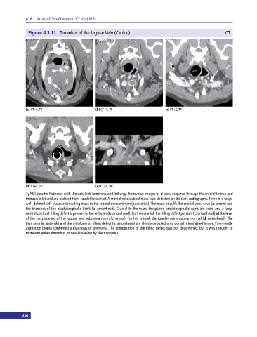
Figure 4.3.11 Thrombus of the Jugular Vein (Canine) CT
(a) CT+C, TP (b) CT+C, TP (c) CT+C, TP
(d) CT+C, TP (e) CT+C, DP
7y FS Labrador Retriever with thoracic limb lameness and lethargy. Transverse images a–d were acquired through the cranial thorax and
thoracic inlet and are ordered from caudal to cranial. A cranial mediastinal mass was detected on thoracic radiographs. There is a large,
well‐defined soft‐tissue attenuating mass in the cranial mediastinum (a: asterisk). The mass engulfs the cranial vena cava (a: arrow) and
the branches of the brachiocephalic trunk (a: arrowhead). Cranial to the mass, the paired brachiocephalic veins are seen, and a large
central contrast filling defect is present in the left vein (b: arrowhead). Further cranial, the filling defect persists (c: arrowhead) at the level
of the convergence of the jugular and subclavian vein (c: arrow). Further cranial, the jugular veins appear normal (d: arrowhead). The
thymoma (e: asterisk) and the intraluminal filling defect (e: arrowhead) are clearly depicted on a dorsal reformatted image. Fine‐needle
aspiration biopsy confirmed a diagnosis of thymoma. The composition of the filling defect was not determined, but it was thought to
represent either thrombus or caval invasion by the thymoma.
416