
Page 431 - Atlas of Small Animal CT and MRI
P. 431
Mediastinum and esophagus 421
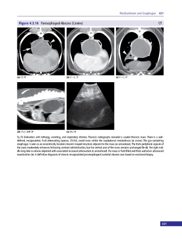
Figure 4.3.16 Paresophageal Abscess (Canine) CT
(a) CT, TP (b) CT+C, TP (c) CT+C, TP
(d) CT+C, MIP, SP (e) US, OP
5y FS Dalmation with lethargy, vomiting, and respiratory distress. Thoracic radiographs revealed a caudal thoracic mass. There is a well‐
defined, encapsulated, fluid‐attenuating (approx. 35 HU), ovoid mass within the caudodorsal mediastinum (a: arrow). The gas‐containing
esophagus is seen as an eccentrically located crescent‐shaped structure adjacent to the mass (a: arrowhead). The thick peripheral capsule of
the mass moderately enhances following contrast administration, but the central part of the mass remains unchanged (b–d). The right mid
dle lung lobe is volume depleted with associated increased attenuation (c: arrowhead). The mass is fluid filled and thick walled on ultrasound
examination (e). A definitive diagnosis of chronic encapsulated paraesophageal bacterial abscess was based on excisional biopsy.
421