
Page 436 - Atlas of Small Animal CT and MRI
P. 436
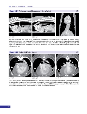
426  Atlas of Small Animal CT and MRI


              Figure 4.4.1  Peritoneopericardial Diaphragmatic Hernia (Feline)                             CT


             (a) CT+C, SP                     (b) CT+C, SP                     (c) CT+C, DP
             Adult  MC  Maine Coon  with  chronic  cough  and  suspected  peritoneopericardial  diaphragmatic  hernia  based  on  previous  thoracic
               radiographs. Images a and b are at slightly different levels in the sagittal plane. Part of the liver is cranially displaced into the pericardial
             sac, causing encroachment on, and cranial displacement of, the heart (a–c: arrow). The defect in the diaphragm (b: arrowheads) and
             characteristic branching hepatic vasculature can be seen (a,c: arrowhead). Echocardiography confirmed the presence of herniated liver
             in the pericardial sac.


              Figure 4.4.2  Pericardial Effusion (Canine)                                                  CT


             (a) CT+C, TP                     (b) CT+C, TP                     (c) CT+C, TP
             11y FS Terrier cross with previously diagnosed pericardial effusion. A moderate volume of pericardial effusion is present, surrounded by
             the uniformly thin, mildly contrast‐enhancing parietal pericardium (a–c: arrowheads). The heterogeneity of the heart is due to contrast‐
             enhanced blood in the cardiac chambers. The average attenuation of pericardial fluid was approximately 12 HU both before and after
             contrast administration. Cytologic analysis revealed the fluid to be a modified transudate.


           426