
Page 437 - Atlas of Small Animal CT and MRI
P. 437
Heart, Pulmonary Vasculature, and Great Vessels 427
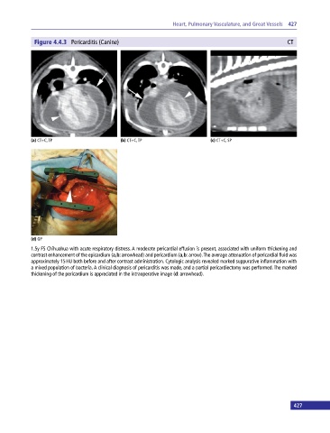
Figure 4.4.3 Pericarditis (Canine) CT
(a) CT+C, TP (b) CT+C, TP (c) CT+C, SP
(d) GP
1.5y FS Chihuahua with acute respiratory distress. A moderate pericardial effusion is present, associated with uniform thickening and
contrast enhancement of the epicardium (a,b: arrowhead) and pericardium (a,b: arrow). The average attenuation of pericardial fluid was
approximately 15 HU both before and after contrast administration. Cytologic analysis revealed marked suppurative inflammation with
a mixed population of bacteria. A clinical diagnosis of pericarditis was made, and a partial pericardiectomy was performed. The marked
thickening of the pericardium is appreciated in the intraoperative image (d: arrowhead).
427