
Page 442 - Atlas of Small Animal CT and MRI
P. 442
432 Atlas of Small Animal CT and MRI
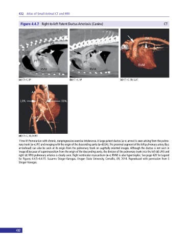
Figure 4.4.7 Right‐to‐left Patent Ductus Arteriosis (Canine) CT
(a) CT+C, SP (b) CT+C, SP (c) CT+C, 3D, LLAT
(d) CT+C, 3D, DORS
11mo M Pomeranian with chronic, nonprogressive exercise intolerance. A large patent ductus (a–c: arrow) is seen arising from the pulmo-
nary trunk (a–c: PT) and merging with the origin of the descending aorta (a–d: DA). The proximal segment of the left pulmonary artery (b,c:
arrowhead) can also be seen at its origin from the pulmonary trunk on sagittally oriented images. Although the ductus is not seen in
image d because of superimposition from the origin of the descending aorta, the division of the pulmonary trunk into the left (d: LPA) and
right (d: RPA) pulmonary arteries is clearly seen. Right ventricular myocardium (a–c: RVM) is also hypertrophic. See page 428 for Legend
for Figures 4.4.5–4.4.15. Susanne Stieger‐Vanegas, Oregon State University, Corvallis, OR, 2014. Reproduced with permission from S
Stieger‐Vanegas.
432