
Page 446 - Atlas of Small Animal CT and MRI
P. 446
436 Atlas of Small Animal CT and MRI
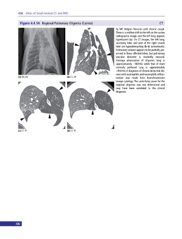
Figure 4.4.14 Regional Pulmonary Oligemia (Canine) CT
9y MC Belgian Tervuren with chronic cough.
There is a midline shift to the left on the survey
radiographic image, and the left lung appears
hyperlucent (a). On CT images, the left lung,
accessory lobe, and part of the right cranial
lobe are hypoattenuating (b–d: arrowheads).
Pulmonary volume appears to be partially pre-
served in these affected lobes, but pulmonary
vascular diameter is markedly reduced.
Average attenuation of oligemic lung is
approximately −960 HU, while that of more
normally perfused lung is approximately
−850 HU. A diagnosis of chronic bronchial dis-
ease with eosinophilic and neutrophilic inflam-
(a) DX, DV (b) CT, DP mation was made from bronchoalveolar
lavage cytology. The underlying cause for the
regional oligemia was not determined and
may have been unrelated to the clinical
diagnosis.
(c) CT, TP (d) CT, TP
436