
Page 449 - Atlas of Small Animal CT and MRI
P. 449
Heart, Pulmonary Vasculature, and Great Vessels 439
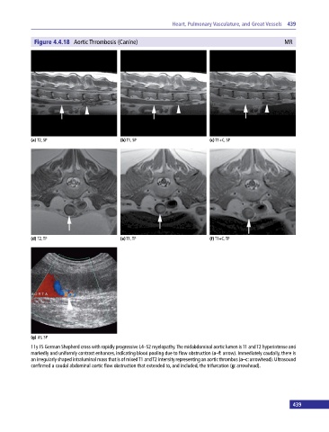
Figure 4.4.18 Aortic Thrombosis (Canine) MR
(a) T2, SP (b) T1, SP (c) T1+C, SP
(d) T2, TP (e) T1, TP (f) T1+C, TP
(g) US, SP
11y FS German Shepherd cross with rapidly progressive L4–S2 myelopathy. The midabdominal aortic lumen is T1 and T2 hyperintense and
markedly and uniformly contrast enhances, indicating blood pooling due to flow obstruction (a–f: arrow). Immediately caudally, there is
an irregularly shaped intraluminal mass that is of mixed T1 and T2 intensity representing an aortic thrombus (a–c: arrowhead). Ultrasound
confirmed a caudal abdominal aortic flow obstruction that extended to, and included, the trifurcation (g: arrowhead).
439