
Page 454 - Atlas of Small Animal CT and MRI
P. 454
444 Atlas of Small Animal CT and MRI
extending into the airway lumen may lead to clinical Degenerative disorders
signs of upper airway obstruction.
CT features of large‐airway neoplasia include focal, Tracheobronchial malacia, a softening of the tracheal
regional, or circumferential thickening of the tracheal cartilages and loss of integrity of the airway walls, is a
wall, and large tumors may appear overtly mass‐like. common cause of large‐airway collapse in people and
The airway patency can be compromised because of has been documented in dogs. Diagnosis is based on a
intraluminal tumor invasion or mural/extramural com greater than 50% collapse of the airway, as observed on
pression. Obstructive bronchial tumors may lead to lobar bronchoscopic examination. Although most veterinary
atelectasis. Tumors usually moderately enhance follow patients are anesthetized or sedated for CT examination
ing contrast medium administration (Figures 4.5.12, and respiration is often assisted, large‐airway collapse is
16
4.5.13, 4.5.14, 4.5.15). sometimes seen (Figure 4.5.16).
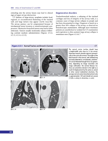
Figure 4.5.1 Normal Trachea and Bronchi (Canine) CT
The normal canine trachea should have
a height:width ratio close to 1.0 (a: arrow).
Mainstem bronchi should originate symmetri
cally at the carina (b: arrows). The origin of the
lobar bronchi in the normal, well‐inflated lung
are easily detected (c: arrowheads), and bron
chi can be followed through five or six genera
tions, depending on image resolution and
image collimation. The normal thoracic tra
chea can sometimes deviate to the right as a
result of displacement by the aorta (c: arrow)
or other cranial mediastinal structures. The
average normal canine bronchial:arterial ratio
is approximately 1.45 and should not exceed
(a) CT, TP (b) CT, TP 2.0 (d: a = artery; b = bronchus; v = vein).
(c) CT, DP (d) CT, TP
444