
Page 456 - Atlas of Small Animal CT and MRI
P. 456
446 Atlas of Small Animal CT and MRI
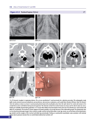
Figure 4.5.3 Tracheal Rupture (Feline) CT
(a) DX, RLAT (b) DX, DV
(c) CT, TP (d) CT, TP (e) CT, TP
(f) CT, DP (g) CT, 3D
11y FS Domestic Longhair in respiratory distress. The cat was anesthetized 1 week previously for a dentistry procedure. The radiographic study
(a,b) reveals extensive pneumomediastinum, pneumothorax, subcutaneous emphysema, and caudal lobar alveolar infiltrates. Near the thoracic
inlet, the trachea is intact (c: arrow). A communicating diverticulum (d: arrowheads) arises from the right side of the trachea (d: arrow) in the
cranial thorax and is well defined during forced inspiration. The diameter of both the tracheal lumen (e: arrow) and the diverticulum (e: arrow
heads) are markedly reduced during expiration. A CT image reformatted in the dorsal plane reveals the size of the diverticulum and tracheal wall
defect (f: arrowheads). The trachea (f: arrow) appears truncated caudally in this image because of the reformatting angle. The intraluminal view
on the virtual bronchoscopic image is from cranial to caudal, and the tracheal bifurcation is seen (g: arrow), as are the lumen of the diverticulum
(g: asterisk) and the margins of the tracheal wall defect (g: arrowheads). Findings on postmortem examination were consistent with tracheal
rupture due to pressure necrosis from an overinflated endotracheal catheter cuff.
446