
Page 458 - Atlas of Small Animal CT and MRI
P. 458
448 Atlas of Small Animal CT and MRI
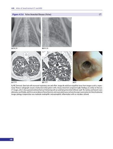
Figure 4.5.6 Feline Bronchial Disease (Feline) CT
(a) DX, DV (b) DX, DV
(c) CT, TP (d) CT, TP (e) ES
6y MC Domestic Shorthair with increased respiratory rate and effort. Images b and d are magnified views from images a and c, respec
tively. Thoracic radiographs reveal a marked bronchial pattern with a heavy interstitial component (a,b). Findings are similar on thoracic
CT images, which show generalized bronchial wall thickening with an underlying interstitial infiltrate (c,d). The trachea and bronchi were
edematous and friable, and there was evidence of bronchiectasis and inspissated mucus on bronchoscopic examination (e). Bronchoalveolar
lavage cytology interpretation was moderate neutrophilic and eosinophilic inflammation with no microbes cultured.
448