
Page 455 - Atlas of Small Animal CT and MRI
P. 455
Airways 445
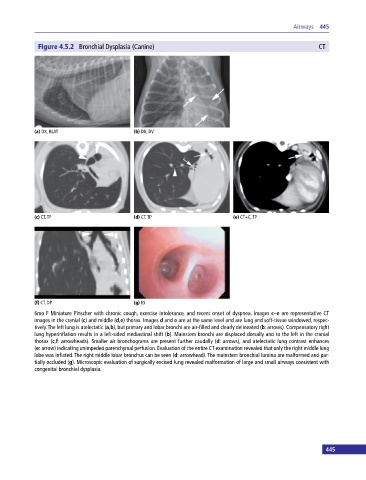
Figure 4.5.2 Bronchial Dysplasia (Canine) CT
(a) DX, RLAT (b) DX, DV
(c) CT, TP (d) CT, TP (e) CT+C, TP
(f) CT, DP (g) ES
6mo F Miniature Pinscher with chronic cough, exercise intolerance, and recent onset of dyspnea. Images c–e are representative CT
images in the cranial (c) and middle (d,e) thorax. Images d and e are at the same level and are lung and soft‐tissue windowed, respec
tively. The left lung is atelectatic (a,b), but primary and lobar bronchi are air‐filled and clearly delineated (b: arrows). Compensatory right
lung hyperinflation results in a left‐sided mediastinal shift (b). Mainstem bronchi are displaced dorsally and to the left in the cranial
thorax (c,f: arrowheads). Smaller air bronchograms are present further caudally (d: arrows), and atelectatic lung contrast enhances
(e: arrow) indicating unimpeded parenchymal perfusion. Evaluation of the entire CT examination revealed that only the right middle lung
lobe was inflated. The right middle lobar bronchus can be seen (d: arrowhead). The mainstem bronchial lumina are malformed and par
tially occluded (g). Microscopic evaluation of surgically excised lung revealed malformation of large and small airways consistent with
congenital bronchial dysplasia.
445