
Page 443 - Atlas of Small Animal CT and MRI
P. 443
Heart, Pulmonary Vasculature, and Great Vessels 433
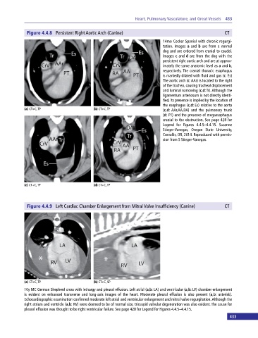
Figure 4.4.8 Persistent Right Aortic Arch (Canine) CT
14mo Cocker Spaniel with chronic regurgi-
tation. Images a and b are from a normal
dog and are ordered from cranial to caudal.
Images c and d are from the dog with the
persistent right aortic arch and are at approx-
imately the same anatomic level as a and b,
respectively. The cranial thoracic esophagus
is markedly dilated with fluid and gas (c: Es)
The aortic arch (c: AAr) is located to the right
of the trachea, causing tracheal displacement
and luminal narrowing (c,d: Tr). Although the
ligamentum arteriosum is not directly identi-
fied, its presence is implied by the location of
the esophagus (c,d: Es) relative to the aorta
(a) CT+C, TP (b) CT+C, TP (c,d: AAr,AA,DA) and the pulmonary trunk
(d: PT) and the presence of megaesophagus
cranial to the obstruction. See page 428 for
Legend for Figures 4.4.5–4.4.15. Susanne
Stieger‐Vanegas, Oregon State University,
Corvallis, OR, 2014. Reproduced with permis-
sion from S Stieger‐Vanegas.
(c) CT+C, TP (d) CT+C, TP
Figure 4.4.9 Left Cardiac Chamber Enlargement from Mitral Valve Insufficiency (Canine) CT
(a) CT+C, TP (b) CT+C, SP
10y MC German Shepherd cross with lethargy and pleural effusion. Left atrial (a,b: LA) and ventricular (a,b: LV) chamber enlargement
is evident on enhanced transverse and long‐axis images of the heart. Moderate pleural effusion is also present (a,b: asterisk).
Echocardiographic examination confirmed moderate left atrial and ventricular enlargement and mitral valve regurgitation. Although the
right atrium and ventricle (a,b: RV) were deemed to be of normal size, tricuspid valvular degeneration was also evident. The cause for
pleural effusion was thought to be right ventricular failure. See page 428 for Legend for Figures 4.4.5–4.4.15.
433