
Page 439 - Atlas of Small Animal CT and MRI
P. 439
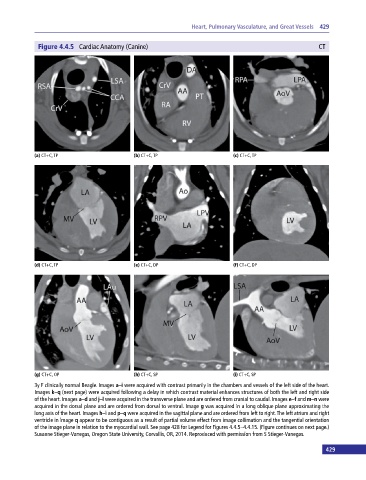
Heart, Pulmonary Vasculature, and Great Vessels  429


              Figure 4.4.5  Cardiac Anatomy (Canine)                                                      CT


            (a) CT+C, TP                     (b) CT+C, TP                     (c) CT+C, TP


            (d) CT+C, TP                     (e) CT+C, DP                     (f) CT+C, DP


            (g) CT+C, OP                     (h) CT+C, SP                     (i) CT+C, SP

            3y F clinically normal Beagle. Images a–i were acquired with contrast primarily in the chambers and vessels of the left side of the heart.
            Images k–q (next page) were acquired following a delay in which contrast material enhances structures of both the left and right side
            of the heart. Images a–d and j–l were acquired in the transverse plane and are ordered from cranial to caudal. Images e–f and m–n were
            acquired in the dorsal plane and are ordered from dorsal to ventral. Image g was acquired in a long oblique plane approximating the
            long axis of the heart. Images h–i and p–q were acquired in the sagittal plane and are ordered from left to right. The left atrium and right
            ventricle in image q appear to be contiguous as a result of partial volume effect from image collimation and the tangential orientation
            of the image plane in relation to the myocardial wall. See page 428 for Legend for Figures 4.4.5–4.4.15. (Figure continues on next page.)
            Susanne Stieger‐Vanegas, Oregon State University, Corvallis, OR, 2014. Reproduced with permission from S Stieger‐Vanegas.

                                                                                                             429