
Page 438 - Atlas of Small Animal CT and MRI
P. 438
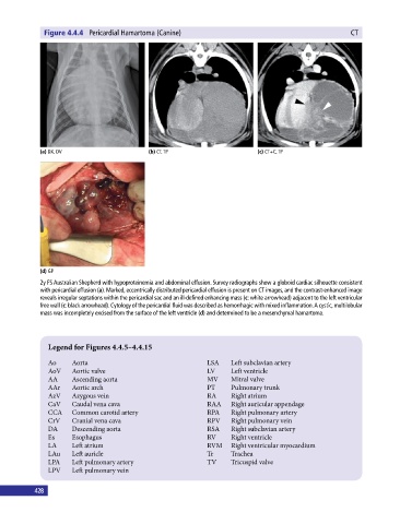
Figure 4.4.4  Pericardial Hamartoma (Canine)                                                 CT


             (a) DX, DV                       (b) CT, TP                       (c) CT+C, TP


             (d) GP

             2y FS Australian Shepherd with hypoproteinemia and abdominal effusion. Survey radiographs show a globoid cardiac silhouette consistent
             with pericardial effusion (a). Marked, eccentrically distributed pericardial effusion is present on CT images, and the contrast‐enhanced image
             reveals irregular septations within the pericardial sac and an ill‐defined enhancing mass (c: white arrowhead) adjacent to the left ventricular
             free wall (c: black arrowhead). Cytology of the pericardial fluid was described as hemorrhagic with mixed inflammation. A cystic, multilobular
             mass was incompletely excised from the surface of the left ventricle (d) and determined to be a mesenchymal hamartoma.


                Legend for Figures 4.4.5–4.4.15


                Ao     Aorta                                    LSA    Left subclavian artery
                AoV    Aortic valve                             LV     Left ventricle
                AA     Ascending aorta                          MV     Mitral valve
                AAr    Aortic arch                              PT     Pulmonary trunk
                AzV    Azygous vein                             RA     Right atrium
                CaV    Caudal vena cava                         RAA    Right auricular appendage
                CCA    Common carotid artery                    RPA    Right pulmonary artery
                CrV    Cranial vena cava                        RPV    Right pulmonary vein
                DA     Descending aorta                         RSA    Right subclavian artery
                Es     Esophagus                                RV     Right ventricle
                LA     Left atrium                              RVM    Right ventricular myocardium
                LAu    Left auricle                             Tr     Trachea
                LPA    Left pulmonary artery                    TV     Tricuspid valve
                LPV    Left pulmonary vein


           428