
Page 441 - Atlas of Small Animal CT and MRI
P. 441
Heart, Pulmonary Vasculature, and Great Vessels 431
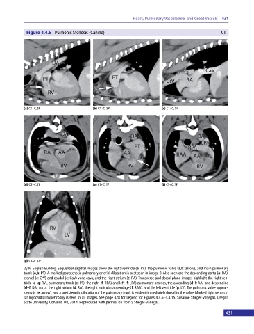
Figure 4.4.6 Pulmonic Stenosis (Canine) CT
(a) CT+C, SP (b) CT+C, SP (c) CT+C, SP
(d) CT+C, TP (e) CT+C, TP (f) CT+C, TP
(g) CT+C, DP
2y M English Bulldog. Sequential sagittal images show the right ventricle (a: RV), the pulmonic valve (a,b: arrow), and main pulmonary
trunk (a,b: PT). A marked poststenotic pulmonary arterial dilatation is best seen in image B. Also seen are the descending aorta (a: DA),
cranial (c: CrV) and caudal (c: CaV) vena cava, and the right atrium (c: RA). Transverse and dorsal plane images highlight the right ven-
tricle (d–g: RV), pulmonary trunk (e: PT), the right (f: RPA) and left (f: LPA) pulmonary arteries, the ascending (d–f: AA) and descending
(d–f: DA) aorta, the right atrium (d: RA), the right auricular appendage (f: RAA), and the left ventricle (g: LV). The pulmonic valve appears
stenotic (e: arrow), and a poststenotic dilatation of the pulmonary trunk is evident immediately dorsal to the valve. Marked right ventricu-
lar myocardial hypertrophy is seen in all images. See page 428 for Legend for Figures 4.4.5–4.4.15. Susanne Stieger‐Vanegas, Oregon
State University, Corvallis, OR, 2014. Reproduced with permission from S Stieger‐Vanegas.
431