
Page 432 - Atlas of Small Animal CT and MRI
P. 432
422 Atlas of Small Animal CT and MRI
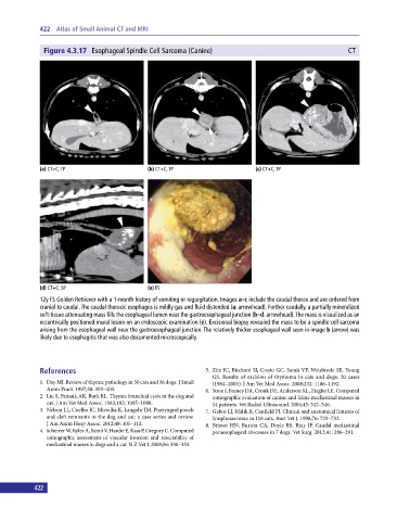
Figure 4.3.17 Esophageal Spindle Cell Sarcoma (Canine) CT
(a) CT+C, TP (b) CT+C, TP (c) CT+C, TP
(d) CT+C, SP (e) ES
12y FS Golden Retriever with a 1‐month history of vomiting or regurgitation. Images a–c include the caudal thorax and are ordered from
cranial to caudal. The caudal thoracic esophagus is mildly gas and fluid distended (a: arrowhead). Further caudally, a partially mineralized
soft‐tissue attenuating mass fills the esophageal lumen near the gastroesophageal junction (b–d: arrowhead). The mass is visualized as an
eccentrically positioned mural lesion on an endoscopic examination (e). Excisional biopsy revealed the mass to be a spindle cell sarcoma
arising from the esophageal wall near the gastroesophageal junction. The relatively thicker esophageal wall seen in image b (arrow) was
likely due to esophagitis that was also documented microscopically.
References 5. Zitz JC, Birchard SJ, Couto GC, Samii VF, Weisbrode SE, Young
GS. Results of excision of thymoma in cats and dogs: 20 cases
1. Day MJ. Review of thymic pathology in 30 cats and 36 dogs. J Small (1984–2005). J Am Vet Med Assoc. 2008;232: 1186–1192.
Anim Pract. 1997;38: 393–403. 6. Yoon J, Feeney DA, Cronk DE, Anderson KL, Ziegler LE. Computed
2. Liu S, Patnaik AK, Burk RL. Thymic branchial cysts in the dog and tomographic evaluation of canine and feline mediastinal masses in
cat. J Am Vet Med Assoc. 1983;182: 1095–1098. 14 patients. Vet Radiol Ultrasound. 2004;45: 542–546.
3. Nelson LL, Coelho JC, Mietelka K, Langohr IM. Pharyngeal pouch 7. Gabor LJ, Malik R, Canfield PJ. Clinical and anatomical features of
and cleft remnants in the dog and cat: a case series and review. lymphosarcoma in 118 cats. Aust Vet J. 1998;76: 725–732.
J Am Anim Hosp Assoc. 2012;48: 105–112. 8. Brissot HN, Burton CA, Doyle RS, Bray JP. Caudal mediastinal
4. Scherrer W, Kyles A, Samii V, Hardie E, Kass P, Gregory C. Computed paraesophageal abscesses in 7 dogs. Vet Surg. 2012;41: 286–291.
tomographic assessment of vascular invasion and resectability of
mediastinal masses in dogs and a cat. N Z Vet J. 2008;56: 330–333.
422