
Page 430 - Atlas of Small Animal CT and MRI
P. 430
420 Atlas of Small Animal CT and MRI
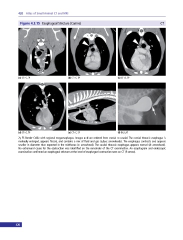
Figure 4.3.15 Esophageal Stricture (Canine) CT
(a) CT+C, TP (b) CT+C, TP (c) CT+C, TP
(d) CT+C, TP (e) CT+C, SP (f) DX, LAT
2y FS Border Collie with regional megaesophagus. Images a–d are ordered from cranial to caudal. The cranial thoracic esophagus is
markedly enlarged, appears flaccid, and contains a mix of fluid and gas (a,b,e: arrowheads). The esophagus contracts and appears
smaller in diameter than expected in the midthorax (c: arrowhead). The caudal thoracic esophagus appears normal (d: arrowhead).
No extramural cause for the obstruction was identified on the remainder of the CT examination. An esophagram and endoscopic
examination confirmed an esophageal stricture at the level of esophageal contraction seen on CT (f: arrow).
420