
Page 425 - Atlas of Small Animal CT and MRI
P. 425
Mediastinum and esophagus 415
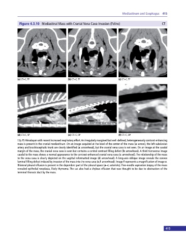
Figure 4.3.10 Mediastinal Mass with Cranial Vena Cava Invasion (Feline) CT
(a) CT+C, TP (b) CT+C, TP (c) CT+C, TP
(d) CT+C, SP (e) CT+C, OP (f) CT+C, OP
12y FS Himalayan with recent increased respiratory effort. An irregularly margined but well‐defined, heterogeneously contrast‐enhancing
mass is present in the cranial mediastinum. On an image acquired at the level of the center of the mass (a: arrow), the left subclavian
artery and brachiocephalic trunk are clearly identified (a: arrowhead), but the cranial vena cava is not seen. On an image at the caudal
margin of the mass, the cranial vena cava is seen but contains a central contrast filling defect (b: arrowhead). A third transverse image
caudal to the mass shows a normal appearance to the contrast‐enhanced cranial vena cava (c: arrowhead). The relationship of the mass
to the vena cava is clearly depicted on the sagittal reformatted image (d: arrowhead). A long‐axis oblique image reveals the convex
luminal filling defect induced by invasion of the mass into the vena cava (e,f: arrowhead). Image f represents a magnification of image e.
Bilateral pleural effusion is present in the dependent part of the pleural space (a–c: asterisks). Fine‐needle aspiration biopsy of the mass
revealed epithelial neoplasia, likely thymoma. The cat also had a chylous effusion that was thought to be due to obstruction of the
terminal thoracic duct by the mass.
415