
Page 424 - Atlas of Small Animal CT and MRI
P. 424
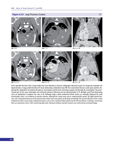
414 Atlas of Small Animal CT and MRI
Figure 4.3.9 Large Thymoma (Canine) CT
(a) CT, TP (b) CT, TP (c) CT+C, DP
(d) CT+C, TP (e) CT+C, TP (f) CT+C, DP
9y FS Labrador Retriever with a large mediastinal mass detected on thoracic radiographs obtained as part of a diagnostic evaluation for
hypercalcemia. A large, predominantly soft‐tissue attenuating mediastinal mass fills the cranioventral thoracic cavity (a,b: asterisk), dis
placing the mediastinal vasculature dorsally (a: arrowheads) and the heart and lungs caudally and dorsally (b: arrowheads). The paren
chymal part of the mass moderately and uniformly enhances following contrast administration (c–f). Multiple small fluid‐attenuating
cysts are distributed throughout the mass (c–f). Although major cranial mediastinal blood vessels are markedly displaced (d: small
arrowheads), there is no evidence of vascular invasion, although the cranial vena cava is compressed (d: arrow). The right internal tho
racic vein is prominent because of increased venous return from mass perfusion (d: large arrowhead). The ventral reflection of the cranial
mediastinum often causes large mediastinal masses such as this to preferentially expand into the left hemithorax, resulting in asymmetry
that can sometimes mimic a left cranial lung lobe mass. Thymoma without vascular invasion was confirmed by excisional biopsy.
414