
Page 429 - Atlas of Small Animal CT and MRI
P. 429
Mediastinum and esophagus 419
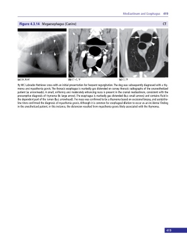
Figure 4.3.14 Megaesophagus (Canine) CT
(a) DX, RLAT (b) CT+C, TP (c) CT, TP
9y MC Labrador Retriever cross with an initial presentation for frequent regurgitation. The dog was subsequently diagnosed with a thy
moma and myasthenia gravis. The thoracic esophagus is markedly gas distended on survey thoracic radiographs of the unanesthetized
patient (a: arrowheads). A small, uniformly and moderately enhancing mass is present in the cranial mediastinum, consistent with the
presumptive diagnosis of thymoma (b: large arrow). The esophagus is markedly gas distended (b,c: small arrows) and contains fluid in
the dependent part of the lumen (b,c: arrowhead). The mass was confirmed to be a thymoma based on excisional biopsy, and acetylcho
line titers confirmed the diagnosis of myasthenia gravis. Although it is common for esophageal dilation to occur as an incidental finding
in the anesthetized patient, in this instance, the distension resulted from myasthenia gravis likely associated with the thymoma.
419