
Page 428 - Atlas of Small Animal CT and MRI
P. 428
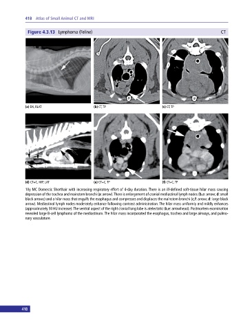
418 Atlas of Small Animal CT and MRI
Figure 4.3.13 Lymphoma (Feline) CT
(a) DX, RLAT (b) CT, TP (c) CT, TP
(d) CT+C, MIP, LAT (e) CT+C, TP (f) CT+C, TP
10y MC Domestic Shorthair with increasing respiratory effort of 4‐day duration. There is an ill‐defined soft‐tissue hilar mass causing
depression of the trachea and mainstem bronchi (a: arrow). There is enlargement of cranial mediastinal lymph nodes (b,e: arrow; d: small
black arrows) and a hilar mass that engulfs the esophagus and compresses and displaces the mainstem bronchi (c,f: arrow; d: large black
arrow). Mediastinal lymph nodes moderately enhance following contrast administration. The hilar mass uniformly and mildly enhances
(approximately 30 HU increase). The ventral aspect of the right cranial lung lobe is atelectatic (b,e: arrowhead). Postmortem examination
revealed large B‐cell lymphoma of the mediastinum. The hilar mass incorporated the esophagus, trachea and large airways, and pulmo
nary vasculature.
418