
Page 427 - Atlas of Small Animal CT and MRI
P. 427
Mediastinum and esophagus 417
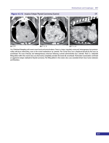
Figure 4.3.12 Invasive Ectopic Thyroid Carcinoma (Canine) CT
(a) CT, TP (b) CT+C, TP (c) CT+C, SP
10y F Shetland Sheepdog with recent onset facial and cervical edema. There is a large, irregularly contoured, heterogeneous but predomi
nately soft‐tissue attenuating, mass in the cranial mediastinum (a: asterisk). The cranial vena cava is displaced dorsally by the mass (a:
arrowhead). The mass intensely and heterogeneously enhances following contrast administration (b,c: asterisk). There is a lobulated
filling defect within the cranial vena cava that nearly completely occludes the lumen (b: arrowhead). Postmortem examination confirmed
an aggressive ectopic mediastinal thyroid carcinoma. The filling defect in the cranial vena cava consisted of both local tumor extension
and thrombus.
417