
Page 416 - Atlas of Small Animal CT and MRI
P. 416
406 Atlas of Small Animal CT and MRI
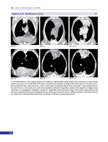
Figure 4.2.10 Mesothelioma (Canine) CT
(a) CT, TP (b) CT, TP (c) CT+C, TP
(d) CT, TP (e) CT, TP (f) CT+C, TP
11y FS Golden Retriever with coughing, dyspnea, and weight loss. Representative images include ventral and dorsal recumbent images
acquired cranial (a–c) and caudal (d–f) to the heart. An ill‐defined mass extends the length of the ventral thorax on the sternal recum-
bent unenhanced images (a,d: asterisk; d: L = liver). A small volume of dependent pleural fluid is also present, which partially obscures
the mass. The mass is more clearly seen on the dorsal recumbent unenhanced images (b,e: asterisk), and incorporation of adipose tissue
contributes to a heterogeneous attenuation caudally (e). Comparable contrast‐enhanced images reveal uniform enhancement of the
soft‐tissue component of the mass (c,f). Dependent pleural fluid is again seen (e,f: arrows). Cytologic evaluation of the pleural effusion
was interpreted as mild mesothelial proliferation. Tissue biopsy of the mass revealed mesothelioma.
406 407