
Page 411 - Atlas of Small Animal CT and MRI
P. 411
Pleural Space 401
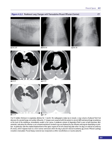
Figure 4.2.2 Positional Lung Changes with Transudative Pleural Effusion (Canine) CT
(a) DX, LAT (b) DX, DV (c) DX, VD
(d) CT, TP (e) CT, TP
(f) CT, TP (g) CT, TP
10y FS Golden Retriever in respiratory distress for 1 month. The radiographic study (a–c) reveals a large volume of pleural fluid that
obscures the aerated lungs and cardiac silhouette. CT images were acquired with the patient in sternal (d,f) and dorsal (e,g) recumbency
at the level of the midthorax, immediately caudal to the carina. A moderate volume of dependent fluid is seen in both positions (d,e:
asterisks), indicating the effusion is not compartmentalized. Aerated lungs are displaced by the effusion and migrate depending on posi-
tion. In particular, note the marked displacement of the apical region of the left cranial lung lobe when the dog is in sternal recumbency
(f: arrow), which migrates back to a more normal orientation when the dog is placed in dorsal recumbency (g: arrow). Effusion cytology
revealed a transudate. Pleural biopsy material was interpreted as either mesothelioma or reactive pleuritis.
400 401