
Page 406 - Atlas of Small Animal CT and MRI
P. 406
396 Atlas of Small Animal CT and MRI
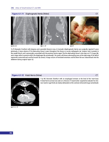
Figure 4.1.11 Diaphragmatic Hernia (Feline) CT
(a) DX, DV (b) CT, DP (c) GP
7y FS Domestic Shorthair with dyspnea and a possible thoracic mass. A traumatic diaphragmatic hernia was surgically repaired 5 years
previously. A large volume of fat attenuating tissue is seen throughout the thorax on survey radiographs (a). Surgical wire is present in
the caudal thorax and is presumably associated with the previous hernia repair. The fat‐attenuating tissue is also seen on CT images (b),
and a large defect is present within the diaphragm (b: arrowheads). The ovoid, soft‐tissue attenuating mass at the right edge of the defect
represents contracted and scarified muscle (b: arrow). A large volume of herniated omentum and falciform fat was reduced back into the
abdomen during surgical repair (c).
Figure 4.1.12 Hiatal Hernia (Feline) CT
1.5y MC Domestic Shorthair with an esophageal stricture at the level of the heart base.
A hiatal hernia (arrow) was seen on a thoracic CT examination acquired to evaluate the stric-
ture. Gastric rugal folds are clearly delineated on this contrast‐enhanced image (arrowhead).
(a) CT+C, TP
396 397