Page 403 - Atlas of Small Animal CT and MRI
P. 403

Thoracic wall and Diaphragm  393


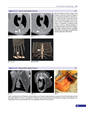
              Figure 4.1.5  Sternal Osteomyelitis (Canine)                                                CT
                                                                             18mo MC Doberman Pinscher. Images a and
                                                                             b represent consecutive transverse images of
                                                                             the cranial thorax ordered from cranial to cau-
                                                                             dal. Unorganized bone destruction involving
                                                                             the second sternabral segment (a–d: arrow-
                                                                             head) is evident. A pathologic fracture is also
                                                                             present (c: arrowhead). A moderate volume
                                                                             of  pleural fluid has collected bilaterally in
                                                                             the dependent pleural space (a,b: asterisks).
                                                                             Bone  biopsy  confirmed  chronic  neutrophilic
                                                                             osteomyelitis, and cytology of the pleural fluid
                                                                             revealed suppurative inflammation.


            (a) CT, TP                       (b) CT, TP


            (c) CT, 3D, LLAT                 (d) CT, 3D, VENT


              Figure 4.1.6  Thoracic Wall Lipoma (Canine)                                                 CT


            (a) CT, DP                       (b) CT, TP                       (c) GP

            9y MC Coonhound with a left thoracic wall and axillary mass. A large, fat‐attenuating mass is present in the left lateral body wall and
            axilla (a,b: asterisk). The mass is bounded by the latissimus dorsi muscle laterally (a,b: arrowhead) and appears homogeneous and
            encapsulated. The mass was removed en bloc (c), and biopsy confirmed it to be a lipoma.


 392                                                                                                         393
   398   399   400   401   402   403   404   405   406   407   408