
Page 375 - Atlas of Small Animal CT and MRI
P. 375
Intervertebral disk disease and other degenerative disorders 365
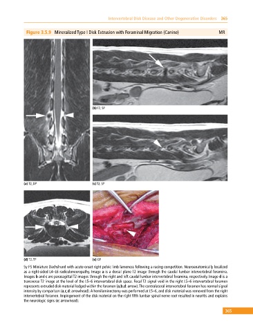
Figure 3.5.9 Mineralized Type I Disk Extrusion with Foraminal Migration (Canine) MR
(b) T2, SP
(a) T2, DP (c) T2, SP
(d) T2, TP (e) GP
5y FS Miniature Dachshund with acute‐onset right pelvic limb lameness following a racing competition. Neuroanatomically localized
as a right‐sided L4–L6 radiculoneuropathy. Image a is a dorsal plane T2 image through the caudal lumbar intervertebral foramina.
Images b and c are parasagittal T2 images through the right and left caudal lumbar intervertebral foramina, respectively. Image d is a
transverse T2 image at the level of the L5–6 intervertebral disk space. Focal T2 signal void in the right L5–6 intervertebral foramen
represents extruded disk material lodged within the foramen (a,b,d: arrow). The contralateral intervertebral foramen has normal signal
intensity by comparison (a,c,d: arrowhead). A hemilaminectomy was performed at L5–6, and disk material was removed from the right
intervertebral foramen. Impingement of the disk material on the right fifth lumbar spinal nerve root resulted in neuritis and explains
the neurologic signs (e: arrowhead).
365