Page 622 - Atlas of Small Animal CT and MRI
P. 622

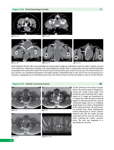
Figure 5.9.8  Penile Hemorrhage (Canine)                                                     CT


             (a) CT+C, TP                     (b) CT+C, TP


             (c) CT+C, TP                     (d) CT+C, TP                     (e) CT+C, TP
             4y MC Doberman Pinscher with a mass extending from rectum to penis. Images are ordered from cranial to caudal. A catheter is present
             in the urethra (a–e: solid arrows), extending to the urinary bladder (a: asterisk). There is a large tubular soft‐tissue and fluid attenuating
             mass parallel to the urethra that extends from the pelvic canal to the penis (b–e: open arrows). The mass does not contrast enhance cen­
             trally and has a rim of peripheral enhancement. Fine‐needle aspirates revealed blood and fat cells, and the mass was presumed to be a
             hematoma. Coagulopathy was not identified; however, there was a history of trauma, and the mass began to reduce in size after 48 hours.


              Figure 5.9.9  Prostatic Carcinoma (Canine)                                                  MR

                                                                              13y MC Leonberger with stranguria and pol­
                                                                              lakiuria. The prostate gland is enlarged (a–c:
                                                                              arrows) with a large cavitary region in the
                                                                              left lobe (b: solid arrowhead) that is hyper­
                                                                              intense on T2 images and hypointense on
                                                                              T1 images. The urethra (b: open arrowhead)
                                                                              appears to communicate with this cavity on
                                                                              unenhanced images and has an irregularly
                                                                              shaped lumen (a–c). There is mineralization
                                                                              of the dorsal parenchyma (b: small arrow).
                                                                              The prostatic mass shows heterogeneous
             (a) T1, TP                       (b) T2, TP
                                                                              contrast enhancement (c: arrow). Contrast‐
                                                                              enhanced urine fills the urethra (d: open
                                                                              arrowhead) and the cavity (d: solid arrow­
                                                                              head),  confirming  the  urethral  communi­
                                                                              cation.  The mass was diagnosed as a
                                                                              transitional cell carcinoma.


             (c) T1+C, TP                     (d) SPGR+C, DP
           612
   617   618   619   620   621   622   623   624   625   626   627