
Page 619 - Atlas of Small Animal CT and MRI
P. 619
Reproductive Tract 609
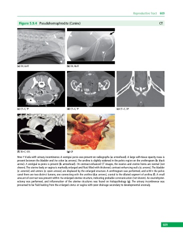
Figure 5.9.4 Pseudohermaphrodite (Canine) CT
(a) DX, LLAT (b) DX, RLAT
(c) CT+C, TP (d) CT+C, TP (e) CT+C, DP
(f) 3D+C, OBL (g) GP
9mo F Viszla with urinary incontinence. A vestigial penis was present on radiographs (a: arrowhead). A large soft‐tissue opacity mass is
present between the bladder and the colon (a: arrows). The urethra is slightly widened in the pelvic region on the urethrogram (b: black
arrow). A vestigial os penis is present (b: arrowhead). On contrast‐enhanced CT images, the ovaries and uterine horns are normal (not
shown). The uterine body or vagina is markedly enlarged and fluid filled with thickened, contrast‐enhancing walls (c: arrows). The bladder
(c: asterisk) and ureters (c: open arrows) are displaced by the enlarged structure. A urethrogram was performed, and within the pelvic
canal there are two distinct lumens, one connecting with the urethra (d,e: arrows), cranial to the dilated segment of urethra (f). A small
amount of contrast was present within the enlarged uterine structure, indicating probable communication (not shown). An ovariohyster
ectomy was performed, and inflammation of the uterine structures was found on histopathology (g). The urinary incontinence was
presumed to be fluid leaking from the enlarged uterus or vagina with poor drainage secondary to developmental anomaly.
609