
Page 616 - Atlas of Small Animal CT and MRI
P. 616
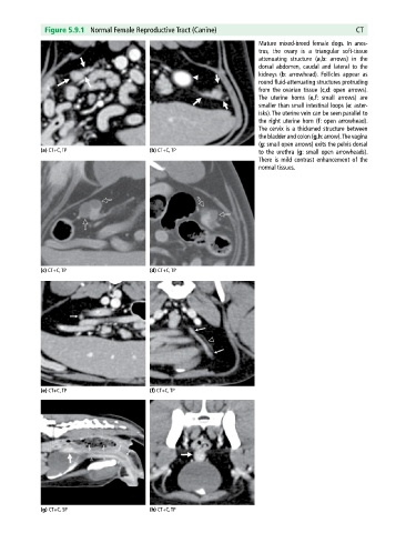
Figure 5.9.1 Normal Female Reproductive Tract (Canine) CT
Mature mixed‐breed female dogs. In anes
trus, the ovary is a triangular soft‐tissue
attenuating structure (a,b: arrows) in the
dorsal abdomen, caudal and lateral to the
kidneys (b: arrowhead). Follicles appear as
round fluid‐attenuating structures protruding
from the ovarian tissue (c,d: open arrows).
The uterine horns (e,f: small arrows) are
smaller than small intestinal loops (e: aster
isks). The uterine vein can be seen parallel to
the right uterine horn (f: open arrowhead).
The cervix is a thickened structure between
the bladder and colon (g,h: arrow). The vagina
(g: small open arrows) exits the pelvis dorsal
(a) CT+C, TP (b) CT+C, TP to the urethra (g: small open arrowheads).
There is mild contrast enhancement of the
normal tissues.
(c) CT+C, TP (d) CT+C, TP
(e) CT+C, TP (f) CT+C, TP
(g) CT+C, SP (h) CT+C, TP