
Page 621 - Atlas of Small Animal CT and MRI
P. 621
Reproductive Tract 611
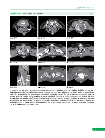
Figure 5.9.7 Paraprostatic Cyst (Canine) CT
(a) CT+C, TP (b) CT+C, TP (c) CT+C, TP
(d) CT+C, TP (e) CT+C, TP (f) CT+C, TP
(g) CT+C, DP (h) CT+C, TP (i) CT+C, TP
2y MC Dachshund with urinary incontinence. Images a–f are ordered from cranial to caudal. There is a large fluid‐filled structure with a
thickened contrast‐enhancing wall on the left side of the caudal abdomen (a–c,g: solid arrow). The urinary bladder (a,g: asterisk) and
urethra (b,c,g: open arrow) also have thickened walls and are displaced to the right. The cyst is continuous, with irregular enhancing
prostatic tissue that contains multiple smaller cysts (c–e,g: arrowhead). The thickened urethra continues through the right side of the
prostate gland and caudal to it (e,f: arrow). The relative positions of the structures are shown on the dorsal reformatted image (g).
A urethrogram was performed, and the paraprostatic cyst filled with contrast medium (h,i: solid arrow). The lumen of the urethra is
moderately irregular indicating urethritis (h,i: open arrows). The cyst was surgically resected with the remnant omentalized. Cystitis and
cyst sepsis were present on microbial culture.
611