
Page 623 - Atlas of Small Animal CT and MRI
P. 623
Reproductive Tract 613
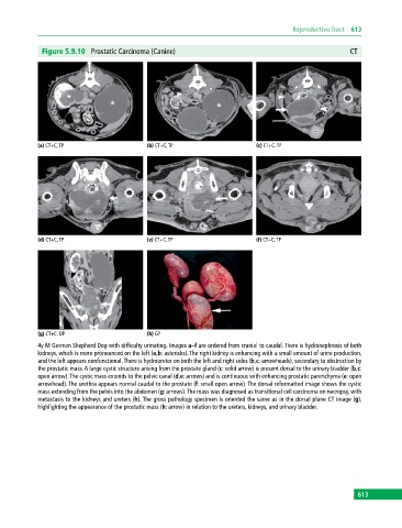
Figure 5.9.10 Prostatic Carcinoma (Canine) CT
(a) CT+C, TP (b) CT+C, TP (c) CT+C, TP
(d) CT+C, TP (e) CT+C, TP (f) CT+C, TP
(g) CT+C, DP (h) GP
4y M German Shepherd Dog with difficulty urinating. Images a–f are ordered from cranial to caudal. There is hydronephrosis of both
kidneys, which is more pronounced on the left (a,b: asterisks). The right kidney is enhancing with a small amount of urine production,
and the left appears nonfunctional. There is hydroureter on both the left and right sides (b,c: arrowheads), secondary to obstruction by
the prostatic mass. A large cystic structure arising from the prostate gland (c: solid arrow) is present dorsal to the urinary bladder (b,c:
open arrow). The cystic mass extends to the pelvic canal (d,e: arrows) and is continuous with enhancing prostatic parenchyma (e: open
arrowhead). The urethra appears normal caudal to the prostate (f: small open arrow). The dorsal reformatted image shows the cystic
mass extending from the pelvis into the abdomen (g: arrows). The mass was diagnosed as transitional cell carcinoma on necropsy, with
metastasis to the kidneys and ureters (h). The gross pathology specimen is oriented the same as in the dorsal plane CT image (g),
highlighting the appearance of the prostatic mass (h: arrow) in relation to the ureters, kidneys, and urinary bladder.
613