
Page 612 - Atlas of Small Animal CT and MRI
P. 612
602 Atlas of Small Animal CT and MRI
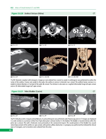
Figure 5.8.20 Urethral Stricture (Feline) CT
(a) CT+C, TP (b) CT+C, TP (c) CT+C, TP
(d) CT+C, SP (e) CT+C, 3D, OBL (f) CT+C, 3D, VENT
11y MC Domestic Longhair with stranguria. Images a–c are ordered from cranial to caudal. A urethrogram was performed to outline the
lumen of the urethra. Cranial and caudal to the pubis, the urethra is normal in diameter (a,c: arrow). The urethral lumen narrows in a
medial to lateral direction at the level of the pubis (b: arrow). The stricture is also seen on a sagittal reformatted image (d: open arrow)
and on 3D reformatted images (e,f: open arrow).
Figure 5.8.21 Pelvic Bladder (Canine) CT
(a) CT+C, SP (b) CT+C, TP (c) CT+C, DP
14y FS Chihuahua with stranguria and pollakiuria. A cystourethrogram was performed under fluoroscopy prior to CT. Images are displayed
in a wide window to counteract artifact from high‐attenuation contrast medium in the bladder. The bladder is located entirely within the
pelvis (a–c: arrows). The wall is thickened, most particularly in the cranioventral aspect. The caudal position of the bladder has caused
lateral displacement of the urethra (c: open arrow), with partial outflow obstruction. A cystopexy was performed that relieved the clinical
signs of stranguria, and no bacteria were cultured from the urine.
602