Page 610 - Atlas of Small Animal CT and MRI
P. 610

600  Atlas of Small Animal CT and MRI


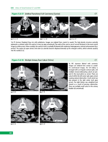
              Figure 5.8.17  Urethral Transitional Cell Carcinoma (Canine)                                 CT


             (a) CT+C, TP                     (b) CT+C, TP                     (c) CT+C, TP
             10y FS German Shepherd Dog mix with pollakiuria. Images are ordered from cranial to caudal. The high‐density structure centrally
               represents a catheter within the urethral lumen (a: open arrowhead). The walls of the urethra are of normal thickness in the most cranial
             image (a: white arrow). More caudally, the urethral wall is markedly thickened with moderate, heterogeneous contrast enhancement (b,c:
             arrows). The vagina (a: open arrow) and colon (a: asterisk) become displaced dorsally by the enlarged urethra, which extends caudally
             into the vestibule (c).


              Figure 5.8.18  Multiple Urinary Tract Calculi (Feline)                                       CT

                                                                              8y MC Japanese Bobtail with azotemia.
                                                                              Images are ordered from cranial to caudal.
                                                                              On  unenhanced images, the left kidney is
                                                                              enlarged with an irregular contour (a,b), and
                                                                              multiple mineral‐attenuating calculi are pre-
                                                                              sent in the renal pelvis (a: arrow). There are
                                                                              calculi within the left ureter (a,b: open arrow)
                                                                              causing ureteral obstruction. Small calculi are
                                                                              also present in the right ureter (c,d: open
                                                                              arrow). The right kidney was atrophied (not
                                                                              shown) as a result of previous obstruction.
                                                                              There are multiple small calculi in the urinary
                                                                              bladder (d: arrowhead).

             (a) CT, TP                       (b) CT, TP


             (c) CT, TP                       (d) CT, TP


           600
   605   606   607   608   609   610   611   612   613   614   615