
Page 606 - Atlas of Small Animal CT and MRI
P. 606
596 Atlas of Small Animal CT and MRI
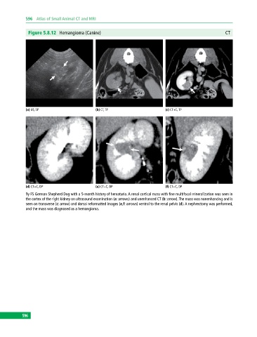
Figure 5.8.12 Hemangioma (Canine) CT
(a) US, SP (b) CT, TP (c) CT+C, TP
(d) CT+C, DP (e) CT+C, DP (f) CT+C, DP
9y FS German Shepherd Dog with a 5‐month history of hematuria. A renal cortical mass with fine multifocal mineralization was seen in
the cortex of the right kidney on ultrasound examination (a: arrows) and unenhanced CT (b: arrow). The mass was nonenhancing and is
seen on transverse (c: arrow) and dorsal reformatted images (e,f: arrows) ventral to the renal pelvis (d). A nephrectomy was performed,
and the mass was diagnosed as a hemangioma.
596