Page 607 - Atlas of Small Animal CT and MRI
P. 607

Urinary Tract  597


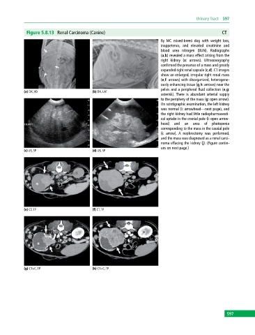
              Figure 5.8.13  Renal Carcinoma (Canine)                                                     CT
                                                                             8y MC mixed‐breed dog with weight loss,
                                                                             inappetence, and elevated creatinine and
                                                                             blood urea nitrogen (BUN). Radiographs
                                                                             (a,b) revealed a mass effect arising from the
                                                                             right kidney (a: arrows). Ultrasonography
                                                                             confirmed the presence of a mass and greatly
                                                                             expanded right renal capsule (c,d). CT images
                                                                             show an enlarged, irregular right renal mass
                                                                             (e,f: arrows) with disorganized, heterogene-
                                                                             ously enhancing tissue (g,h: arrows) near the
                                                                             pelvis and a peripheral fluid collection (e,g:
            (a) DX, VD                       (b) DX, LAT
                                                                             asterisk).  There is abundant arterial supply
                                                                             to the periphery of the mass (g: open arrow).
                                                                             On scintigraphic examination, the left kidney
                                                                             was normal (i: arrowhead—next page), and
                                                                             the right kidney had little radiopharmaceuti-
                                                                             cal uptake in the cranial pole (i: open arrow-
                                                                             head)  and an area of photopenia
                                                                             corresponding to the mass in the caudal pole
                                                                             (i: arrow).  A nephrectomy was performed,
                                                                             and the mass was diagnosed as a renal carci-
                                                                             noma effacing the kidney (j). (Figure contin-
                                                                             ues on next page.)
            (c) US, SP                       (d) US, SP


            (e) CT, TP                       (f) CT, TP


            (g) CT+C, TP                     (h) CT+C, TP


                                                                                                             597
   602   603   604   605   606   607   608   609   610   611   612