
Page 603 - Atlas of Small Animal CT and MRI
P. 603
Urinary Tract 593
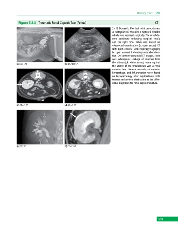
Figure 5.8.8 Traumatic Renal Capsule Tear (Feline) CT
2y FS Domestic Shorthair with uroabdomen.
A cystogram (a) revealed a ruptured bladder,
which was repaired surgically. The uroabdo-
men continued following surgical repair,
and the right renal pelvis was dilated on
ultrasound examination (b: open arrow), CT
(d,f: open arrows), and nephropyelography
(e: open arrows), indicating ureteral obstruc-
tion. On contrast‐enhanced CT images, there
was subcapsular leakage of contrast from
the kidney (c,f: white arrow), revealing that
(a) DX, LAT (b) US, MIP, SP
the source of the uroabdomen was a renal
capsular tear. Ureteral necrosis, subcapsular
hemorrhage, and inflammation were found
on histopathology after nephrectomy, with
trauma and ureteral obstruction as the differ-
ential diagnoses for renal capsular rupture.
(c) CT+C, TP (d) CT+C, TP
(e) DX, VD (f) CT+C, DP
593