
Page 600 - Atlas of Small Animal CT and MRI
P. 600
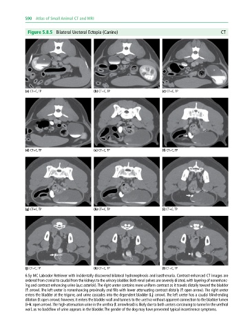
590  Atlas of Small Animal CT and MRI


              Figure 5.8.5  Bilateral Ureteral Ectopia (Canine)                                            CT


             (a) CT+C, TP                     (b) CT+C, TP                     (c) CT+C, TP


             (d) CT+C, TP                     (e) CT+C, TP                     (f) CT+C, TP


             (g) CT+C, TP                     (h) CT+C, TP                     (i) CT+C, TP


             (j) CT+C, TP                     (k) CT+C, TP                     (l) CT+C, TP
             6.5y MC Labrador Retriever with incidentally discovered bilateral hydronephrosis and isosthenuria. Contrast‐enhanced CT images are
             ordered from cranial to caudal from the kidneys to the urinary bladder. Both renal pelves are severely dilated, with layering of nonenhanc-
             ing and contrast‐enhancing urine (a,c: asterisk). The right ureter contains more uniform contrast as it travels distally toward the bladder
             (f: arrow). The left ureter is nonenhancing proximally and fills with lower attenuating contrast distally (f: open arrow). The right ureter
             enters the bladder at the trigone, and urine cascades into the dependent bladder (i,j: arrow). The left ureter has a caudal blind‐ending
             dilation (l: open arrow); however, it enters the bladder wall and tunnels to the urethra without apparent connection to the bladder lumen
             (i–k: open arrow). The high‐attenuation urine in the urethra (l: arrowhead) is likely due to both ureters continuing to tunnel in the urethral
             wall, as no backflow of urine appears in the bladder. The gender of the dog may have prevented typical incontinence symptoms.