
Page 602 - Atlas of Small Animal CT and MRI
P. 602
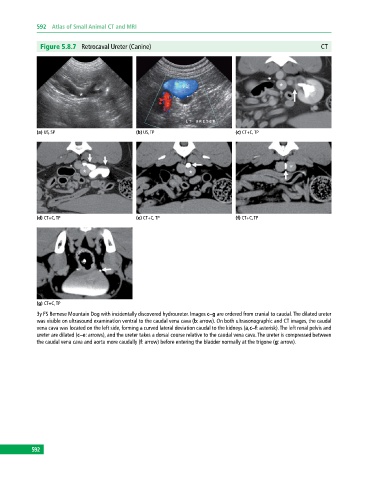
592  Atlas of Small Animal CT and MRI


              Figure 5.8.7  Retrocaval Ureter (Canine)                                                     CT


             (a) US, SP                       (b) US, TP                       (c) CT+C, TP


             (d) CT+C, TP                     (e) CT+C, TP                     (f) CT+C, TP


             (g) CT+C, TP
             3y FS Bernese Mountain Dog with incidentally discovered hydroureter. Images c–g are ordered from cranial to caudal. The dilated ureter
             was visible on ultrasound examination ventral to the caudal vena cava (b: arrow). On both ultrasonographic and CT images, the caudal
             vena cava was located on the left side, forming a curved lateral deviation caudal to the kidneys (a,c–f: asterisk). The left renal pelvis and
             ureter are dilated (c–e: arrows), and the ureter takes a dorsal course relative to the caudal vena cava. The ureter is compressed between
             the caudal vena cava and aorta more caudally (f: arrow) before entering the bladder normally at the trigone (g: arrow).


           592