
Page 598 - Atlas of Small Animal CT and MRI
P. 598
588 Atlas of Small Animal CT and MRI
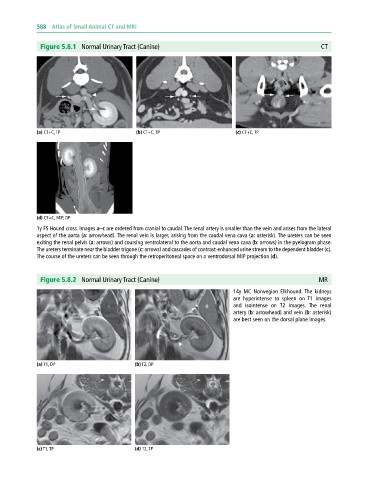
Figure 5.8.1 Normal Urinary Tract (Canine) CT
(a) CT+C, TP (b) CT+C, TP (c) CT+C, TP
(d) CT+C, MIP, DP
1y FS Hound cross. Images a–c are ordered from cranial to caudal. The renal artery is smaller than the vein and arises from the lateral
aspect of the aorta (a: arrowhead). The renal vein is larger, arising from the caudal vena cava (a: asterisk). The ureters can be seen
exiting the renal pelvis (a: arrows) and coursing ventrolateral to the aorta and caudal vena cava (b: arrows) in the pyelogram phase.
The ureters terminate near the bladder trigone (c: arrows) and cascades of contrast‐enhanced urine stream to the dependent bladder (c).
The course of the ureters can be seen through the retroperitoneal space on a ventrodorsal MIP projection (d).
Figure 5.8.2 Normal Urinary Tract (Canine) MR
14y MC Norwegian Elkhound. The kidneys
are hyperintense to spleen on T1 images
and isointense on T2 images. The renal
artery (b: arrowhead) and vein (b: asterisk)
are best seen on the dorsal plane images.
(a) T1, DP (b) T2, DP
(c) T1, TP (d) T2, TP