
Page 608 - Atlas of Small Animal CT and MRI
P. 608
598 Atlas of Small Animal CT and MRI
Figure 5.8.13 (Continued ) CT
(i) NM, DORS (j) GP
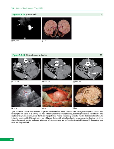
Figure 5.8.14 Nephroblastoma (Canine) CT
(a) CT+C, TP (b) CT+C, TP (c) CT+C, TP
(d) US, SP (e) GP (f) GP
2y MC Doberman Pinscher with hematuria. Images a–c are ordered from cranial to caudal. There is a large, heterogeneous, cavitary mass
replacing the left kidney (a–c: arrows). The mass is heterogeneously contrast enhancing, and urine production is present in the most
caudal cavitary region (c: arrowhead). The CT scan was performed in dorsal recumbency, hence the inverted fluid‐contrast interface. The
left ureter is not identified. The right kidney has mild pelvic dilation with a thin lateral cortex (a: open arrow) and cortical infarct (not
shown). The mass is vascular on Doppler ultrasound (d). A nephrectomy was performed and nephroblastoma with disorganized renal
tissue was diagnosed (e,f).
598
598